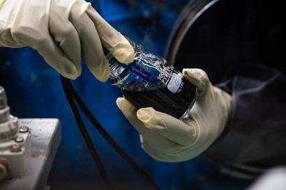
When contemplating methods to sustainably generate environmentally pleasant merchandise, micro organism may not instantly spring to thoughts.
Nevertheless, in recent times scientists have created microbe-semiconductor biohybrids that merge the biosynthetic energy of residing programs with the flexibility of semiconductors to reap mild. These microorganisms use photo voltaic power to transform carbon dioxide into value-added chemical merchandise, equivalent to bioplastics and biofuels. However how that power transport happens in such a tiny, complicated system, and whether or not the method might be improved, remains to be unclear.
A scanning electron microscopy picture exhibits bacterium Ralstonia eutropha cells on prime of a bismuth vanadate semiconductor.
Cornell researchers have developed a multimodal platform to picture these biohybrids with single-cell decision, to higher perceive how they operate and the way they are often optimized for extra environment friendly power conversion.
The group’s paper, “Single-Cell Multimodal Imaging Uncovers Power Conversion Pathways in Biohybrids,” revealed July 27 in Nature Chemistry. The co-lead authors are postdoctoral researcher Bing Fu and former postdoctoral researcher Xianwen Mao.
The challenge was led by Peng Chen, the Peter J.W. Debye Professor of Chemistry within the School of Arts and Sciences. The trouble is an offshoot of a bigger collaboration – with Tobias Hanrath, professor on the Smith College of Chemical and Biomolecular Engineering in Cornell Engineering, and Buz Barstow, Ph.D. ’09, assistant professor of organic and environmental engineering within the School of Agriculture and Life Sciences – that was funded by the U.S. Division of Power (DOE) to discover microscopic imaging of microbes as a strategy to advance bioenergy analysis.
Biohybrid analysis has usually been performed with micro organism in bulk – basically a considerable amount of cells in a bucket, Peng mentioned – emphasizing the general yield of the value-added chemical substances and the collective behaviors of the cells, slightly than the underlying mechanism that permits the complicated chemical transformation.
“Biology may be very heterogeneous. The person cells are very totally different. Now, as a way to interrogate it higher, you actually need to measure it at a single-cell degree,” Chen mentioned. “That is the place we are available. We offer quantitative assessments of protein behaviors and likewise a mechanistic understanding of how the electron transport happens from the semiconductor to the micro organism cell.”
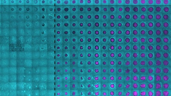
The brand new platform mixed multi-channel fluorescence imaging with photoelectrochemical present mapping to survey the bacterium Ralstonia eutropha. The platform was capable of concurrently picture, monitor and quantitate a number of proteins within the cell whereas additionally measuring the stream of electrons, finally correlating the mobile protein properties and electron transport processes.
The researchers efficiently differentiated the purposeful roles of two forms of hydrogenases – one sure to the cell’s membrane, and a soluble one within the cytoplasm – that assist metabolize hydrogen and drive CO2 fixation. Whereas the soluble hydrogenase is understood to be essential for metabolizing hydrogen, the researchers discovered that the membrane-bound hydrogenase, whereas much less necessary, really facilitates the method and makes it extra environment friendly.
As well as, the researchers obtained the primary experimental proof that the micro organism can uptake a considerable amount of electrons from semiconductor photocatalysts. The group measured the electron present and located it’s three orders of magnitude bigger than what scientists beforehand thought, which means that future micro organism strains may very well be engineered to enhance the effectivity of power conversion.
The researchers additionally found that membrane-bound and soluble hydrogenases play an necessary function in mediating the electron transport from the semiconductor into the cell. In the meantime, not solely can the cell settle for electrons; it may additionally spit them out in the wrong way, with out the help of hydrogenases.
The imaging platform is generalizable sufficient that it may be used to review different biological-inorganic programs, together with yeast, and for different processes, equivalent to nitrogen fixation and pollutant removing.
“Our multimodal imaging platform is highly effective, nevertheless it in fact has its personal limits,” Chen mentioned. “We will picture and examine proteins, however our strategy doesn’t enable us to investigate small molecule compositions. And so one can take into consideration additional integrating our strategy with different strategies – for instance, nanoscale mass spectrometry – so it will be actually highly effective. We’re not there but.”
Co-authors embrace Hanrath and Barstow; postdoctoral researcher Youngchan Park; doctoral college students Zhiheng Zhao, Tianlei Yan, Farshid Salimijazi, and Mokshin Suri; Danielle H. Francis, M.S. ’17; Gained Jung, Ph.D. ’18; former postdoctoral researcher Wenjie Li; and laboratory technician Brooke Pian ’13.
The analysis was supported by the DOE’s Biomolecular Characterization and Imaging Science program.
The researchers madeuse of the Cornell Middle for Supplies Analysis Shared Services, which is supported by means of the Nationwide Science Basis’s MRSEC program.